
Die Dreigliederung der Haut
Export Article Citation as
- Download price : €6
Abstract:
The threefoldness of the skin
If the three layers of the human skin are analysed according to a functional view, a picture of the effective formative forces in the skin is produced.
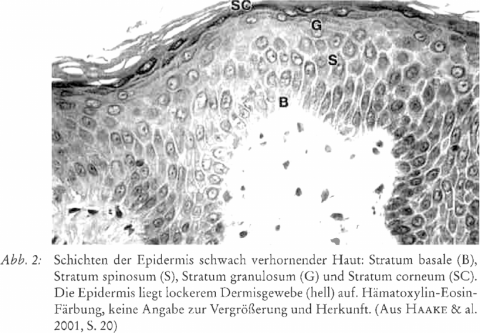
The primary formative force of the outermost layer (epidermis) produces the horny substance which isolates the physical organism from its environment. The production of the horny substance takes place by a death process of the horn-producing cells involved. This is called terminal differentiation. This process of terminal differentiation leads the substance to solidification and rigidity from which it cannot be reintegrated into the living organism but is expelled. Physiologically and biochemically the epidermis shows the characteristics of organs with ectoderrnal origin: the brain and the nerve system.
The internal layer of the skin, the fatty tissue, presents a functional character that is polar to the epidermis. The subcutaneous fatty tissue is part of the metabolic system of the organism. As a store for fats it is situath between uptake of nutritional fats and their release according to the needs of the organism. Moreover, the fatty tissue is an endocrine organ whose horrnoncs affect various metabolic processes. In its lack of rigid, solid structures and the resistance to apoptosis (programmed cell death) of the fat cells it represents a polarity to the epidermis.
The dermis as the middle layer reveals properties of both formative principles. The histological picture of the dermis is determined by the intercellular substance which consists of a structured part, the fibres, and a structureless part, the amorphous matrix. As a tissue the dermis is rich in blood and lymph vessels, and also the intercellular substance is functionally part of the organism’s circulation processes. In the dermis the functions of the rhythmic system predominate.
Thus the skin with its three layers represents all three parts of the whole human organism: nerve-sense system, rhythmic system and digestive system.
References
- BENNINGHOFF, A. & DRENCKHAHN, D. (2004): Anatomie Bd. 2. München
- BRAUN-FALCO, O., PLEWIG, G. & WOLFF, H. II. (1996): Dermatologie und Venetologie, S. 914. Berlin
- CHRISTEN, Ph. & JAUSSI, R. (2005): Biochemie. Berlin
- ELIAS, P. M. & FEINGOLD, K. R. (1992): Lipids and the epidermal water barrier: metabolism‚ regulation, and pathophysiology. Seminars in Dermatology 11 (2): 176 - 182
- FRAYN, K. N. & al. (2003): Integrative physiology of human adipose tissue. International Journal of Obesity 27: 875 - 888
- FREINKEL, R. K. (2001): Metabolism of the skin. In: Freinkel, R. K. & Woodley, D. T. (Eds.), The Biology of the Skin, pp. 191 - 199. New York
- FRENTZ, G. & al. (1991): On circadian rhythms in human epidermal cell proliferation. Acta Dermatologica Venerologica (Stockholm) 71: 85 - 87
- FRITSCH, P. (2004): Dermatologie Venerologie. Berlin
- FUCHS, E. (1990): Epidermal differentiation: The bare essentials. The Journal of Cell Biology 111: 2807 - 2814
- GOLENHOFEN, K. (2000): Physiologie. München
- GRUNDY, S. M. & al. (2004): Definition of metabolic syndrome. Circulation 109: 433 - 438
- HAAKE, A., SCOTT, G. A. & HOLBROOK, K. A. (2001): Structure and function of the skin: overview of the epidermis and dermis. In: Freinkel, R. K. & Woodley D. T. (Eds.)‚ The Biology of the Skin, pp. 19 - 45. New York
- HAUCK, G. (1994): Kapilläre Permeabilität und Mikro-Lymphdrainage. VASA 23: 93 .. 96
- HERRMANN, F., IPPEN, H., SCHAEFER‚ H. & STÜTTGEN, G. (1973): Biochemie der Haut. Stuttgart
- HOLBROOK, K. A. & HOFF, M. S. (1984): Structure of the developing human embryonic and fetal skin. Seminars in Dermatology 3: 185 - 202
- JACHENS, L. (2004): Über die Wirkung von Dermatodoron. Der Merkurstab 57 (6): 408 - 413
- JUREVICS, H. A. & MORELL, P. (1994): Sources of cholesterol for kidney and nerve during development. journal of Lipid Research 35: 112 - 120
- KERSHAW, E. E. & FLIER, J. S. (2004): Adipose tissue as an endocrine organ. The Journal of Clinical Endocrinology & Metabolism 89 (6): 2548 - 2556
- KRSTIC, R. V. (1978): Die Gewebe des Menschen und der Säugetiere. Berlin
- KUBIK, S. (2002): Anatomie des Lymphgefäßsystems. In: Földi, M. & Kubik, S. (Hrsg.), Lehrbuch der Lymphologie, 5. Auflage. München
- LAUE, H. B. VON (1977): Die Formprozesse in Entwicklung und Krankheit der Hautorgane. Stuttgart
- LIEM, K., BEMIS, W. E., WALKER jr., W. F. & GRANDE, L. (2001): Functional Anatomy of the Vertebrates, 3rd edition. Fort Worth
- LINNEMANN, M. & KÜI-IL, M. (2005): Biochemie für Mediziner. Berlin
- LOFFREDA, S. & al. (1998): Leptin regulates proinflammatory immune responses. FASEB Journal 12: 57 - 65
- LORD, G. M. & al. (1998): Leptin modulates the T-cell immune r65ponse and reverses starvation-induced immunosuppression. Nature 394: 897 - 901
- MADISON, K. C. (2003): Batrier function of the skin:»La Raison d'Etre«of the epidermis. The Journal of Investigative Dermatology 121: 231 - 241
- MAGERSTÄDT, K. & GRÄFLIN, G. (1986): Die Haut und ihre Erkrankungen. In: Wolff, 0. (Hrsg.), Das Bild des Menschen als Grundlage der Heilkunst, Bd. 3, S. 502 520. Stuttgart
- MELINO, G. & al. (1998): The cornificd cnvelope: A model of cell death in the skin. In: Kumar, S. (Ed.), Results and Problems in Cell Differentiation, pp. 175 - 212. Berlin
- NEUBERT, R., WOHLRAB, W. & MARSCH, W. (2001): Dermatopharmazie. Stuttgart
- NORDLUND, J. J. & BOISSY, R. E. (2001): The biology of melanocytes. In: Freinkel, R. K. & Woodley, D. T. (Eds.), The Biology of the Skin, pp. 113 - 131. New York
- PONEC, M. & al. (1983): Cultured human skin fibroblasts and keratinocytes: differences in the regulation of cholcsterol synthesis. The Journal of1nvestigative Dermatology 81: 125 - 130
- RYAN, T. J. (1989): Structure and function of lymphatics. The Journal of Investigative Dermatology 93: 185 - 24S
- SCHEFFLER, A. (2001): Heilpflanzcnerkenntnis aus der Sicht der anthroposophischen Medizin am Beispiel der Mistel Viscum album L. In: Scheer, R. & al. (Hrsg.), Die Mistel in der Tumortherapie. Grundlagenforschung und Klinik, S. 519 - 558. Essen
- SCHEFFLER, A. & al. (2004): Zur Heilprozessidee von Birkenrinde und Hautkrankheiten. Der Merkurstah 57 (6): 453 - 466
- SEIDELIN, K. (1995): Patty acid composition of adipose tissue in humans. Implications for the dietary fat-serum cholestcrol-CHD issue. Progress in Lipid Research 34 (3): 199 - 217
- SMITH, L. T. & al. (1982): Structure of the dermal matrix during development and in the adult. The Journal of Investigative Dermatology 79: 935 - 1045
- SOBOTTA,J. & WELSCH, U. (2002): Atlas Histologie. München
- SORISKY, A. & al. (2000): Adiposc cell apoptosis: death in the energy depot. International Journal of Obesity 24 Suppl. 4: S3 - S7
- STEINHOFF, M. & al. (2003): Modern aspects of cutaneous neurogenic inflammation. Archives of Dermatology 139: 1479 - 1488
- STEVENS, A. & LOWE, J. (1997): Histologie des Menschen. Weinheim
- WERTZ, Ph. W. (1992): Epidermal lipids. Seminars in Dermatology 11 (2): 106 - 113
- WERTZ, Ph. W. (2000): Lipids and barrier function of the skin. Acta Dermatologica Venereologica Suppl. 208: 7 - 11
- WILLIAMS, A. C. (2003): Transdermal and topical drug delivery, p. 9. London
- WOLFF, O. (1998): Grundlagen einer geisteswissenschaftlich erweiterten Biochemie, S. 138 ff. Stuttgart
- YOSIPOVICS, G. & al. (1998): 'Time-dependent variations of the skin barrier function in humans: Transepidermal water loss, stratum corneum hydration, skin surface pH, and skin temperature. The Journal of Investigative Dermatology 110: 20 - 23
- ZIBOH, V. A. & CHAPKIN, R. S. (1988): Metabolism and function of skin lipids. Progress in Lipid Research 27: 81 - 105