
Lebendige Mineralwelt im Knochen
Export Article Citation as
- Download price : €6
Abstract:
The mineral realm in living bone
The mineralisation of the skeleton is based on the dissolved calcium, phosphate and carbonate of body fluids and even on the water and carbon dioxide of the earth‘s hydrosphere and atmosphere. But it is also related to the mineral realm‘s crystalline calcium phosphate and carbonate, especially to apatite. The polar extremes of skeleton formation actually lie in the ‚environment‘ which expresses itself in a distinct tendency to two-dimensionality:
- First of all organic-mineral germs arise on and in the outer membranes of the mitochondria of cartilage and bone cells and the outer membranes of extracellular vesicles.
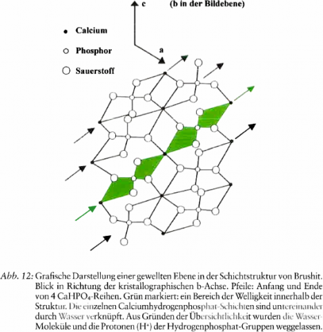
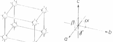
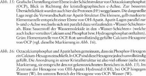
- Apatite precursors (brushite, octacalcium phosphate-like phases) form lamellar structures in close relation to the aqueous realm.
- Collagen fibres gradually become completely coated with biological apatite.
- Despite its three dimensional, networked, stable scaffolding-structure the biological apatite remains connected With two-dimensionality. Its microcrystallinity provides the skeleton with an enormous inner surface area which is in intense exchange with the environment via the hydrate envelopes of the crystallite.
- Non-apatite areas of the surface of the apatite crystallite contain highly reactive, exehangeable, labile phosphate and labile carbonate, which is greatest in the cartilaginous and young bone mineral. Labile carbonate is physiologically decisive for this.
As the skeleton matures stable phosphate and stable carbonate increase. Above all, stable carbonate replaces stable phosphate, the most stable position in the crystal structure. To a lesser extent it is replaced by the more readily exchangeable hydroxide, fluoride or chloride. The latter and the equally exchangeable calcium are localised in particular ,migration zones‘ of the crystal structure.
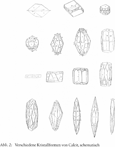
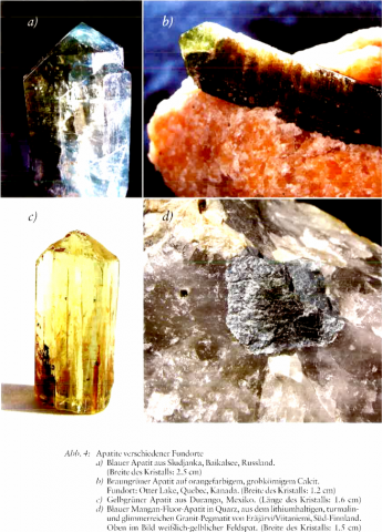
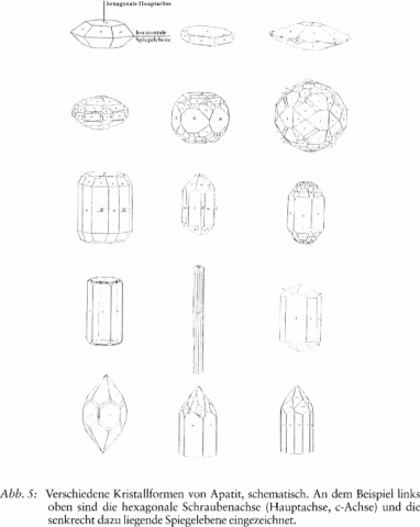
The extent of variation and readiness to partake in processes of apatite are particularly strongly connected with carbonate. Even in the mineral realm, apatite shows an extraordinary capacity for absorption various exogenous ions. Apatite‘s natural typical structural variability makes it particularly suited to its function in the organism. But even the carbonate of the mineral realm is very inclined to variation, between, on the one hand, totally dissolved calcium hydrogen carbonate and, on the other hand, calcite, the mineral with the greatest number of forms.
Something of the nature of bone already resides in the apatite of the mineral realm. No mineral other than apatite can form the skeleton in such a living way, and keep it in connection with life.


References
- ANDERSON, H. C. (1976): Matrix vesicle calcification. Federation Proceedings 35(2): 105-108
- ARNOLD, W H. (2006): Mikromorphologie und Molekularbiologie der Dentinogenese und Amelogenese. Deutsche Zahnärztliche Zeitschrift 61: 524-534
- BACQUET, G., TRUONG, V. Q., VIGNOLES, M., BONEL, G. (1981): EPR detection of acetate ions trapping in B-type carbonated fluorapatites. Journal of Solid State Chemistry 39: 148-153
- BAUER, G. C. H., CARLSSON, A., LINDQUIST, B. (1961): Metabolism and homeostatic function of bone. In: Comar, C. L., Bronner, F., Mineral Metabolism. An Advanced Treatise, Vol. I/B, pp. 607-676. New York, London
- BENESCH, F. (1990): Der Turmalin. Eine Monographie. Stuttgart
- BERNARDI, G. ( 1975): Interactions betwen hydroxyapatite and biological macromolecules (proteins, nucleic acids). Colloques Internationaux du Centre National de la Recherche Scientifique 230: Physico-Chimie et Cristallographie des Apatites d’Interét Biologique, pp. 463-465. Paris
- BETECHTIN, A. G. (1977): Lehrbuch der speziellen Mineralogie, 7. Aufl. (1964), unveränderter Nachdruck (1977). Leipzig
- BIGI, A., COJAZZI, G., PANZAVOLTA, S., RIPAMONTI, A., ROVERI, N., ROMANELLO, M., NORIS SUAREZ, K., MORO, L. (1997): Chemical and structural characterization of the mineral phase from cortical and trabecular bone. Inorganic Biochemistry 6 8(1): 45-51
- BONUCCI, E. (1975): The organicinorganic relationships in calcified organic matrices. Colloques Internationaux du Centre National de la Recherche Scientifique 230: Physico-Chimie et Cristallographie des Apatites d’Interét Biologique, pp. 231-246. Paris
- BRIGHTON, C. T., HUNT, R. M. (1976): Histochemical localization of calcium in growth plate mitochondria and matrix vesicles. Federation Proceedings 35 (2): 143-147
- BUSHINSKY, D. A., SMITH, S. B., GAVRILOV, K. L., GAVRILOV, L. F., LI, J., LEVI-SETTI, R. (2003): Chronic acidosis-induced alteration in bone bicarbonate and phosphate. American Journal of Physiology/Renal Physiology 285 : F532-F539
- CIFUENTES, I., GONZÁLEZ-DÍAZ, P. F., CIFUENTES-DELATTE, L. (1980): Is there a »citrate-apatite« in biological calcified systems? Calcified Tissue International 31(1): 147-151
- CHO, G., WU, Y., ACKERMAN, I. (2003): Detection of hydroxyl ions in bone mineral using solid stage NMR spectroscopy. Science 300: 1123-1127
- CRANE, N. J., POPESCU, V., MORRIS, M. D., STEENHUIS, P., IGNELZI JR., M. A. (2006): Raman spectroscopic evidence for octacalcium phosphate and other transient mineral species deposited during intramembraneous mineralization. Bone: Cell and Molecular Biology, Pathophysiology, Treatment 39: 434-442
- EANES, E. D. (1979): Enamel-apatite: chemistry, structure and properties. Journal of Dental Research 58(3): 829-834
- EDMUNDS, M., SMEDLEY, P. (2005): Fluoride in natural waters. In: Selinus, O., Alloway, B. J., Centento, J. A., Finkelman, R. B., Fuge, R., Lindh, U., Smedley, P. (Eds.), Essentials of Medical Geology. Impacts of the Natural Environment on Public Health, pp. 301-329. Amsterdam, Boston, Heidelberg u. a.
- ELLIOTT, J. C. (1973): The problems of the composition and structure of the mineral components of the hard tissues. Clinical Orthopaedics and Related Research 93: 313-345
- ELLIOT, J. C. (1994): Structure and Chemistry of the Apatites and Other Calcium Orthoposphates. Studies in Inorganic Chemistry 18. Amsterdam, London, New York, Tokyo
- ELLIOT, J. C. (2002): Calcium phosphate biominerals. In: Kohn, M. J., Rakovan, J., Hughes, J. M. (Eds.), Phosphates: Geochemical, Geobiological, and Materials Importance. Reviews in Mineralogy and Geochemistry Vol. 48: 427-453. Washington
- ELLIOT, J. C.; WILSON, R. M.; DOWKER, S. E. P. (2002): Apatite structures. JCDS-International Centre for Diffraction Data, Advances in X-Ray Analysis 45: 172-181
- FLEISCH, H. (1980): Mechanism of calcification. In: Massry, S. G.; Ritz, E.; Jahn, H.; Posphate and Minerals in Health and Disease, pp. 563-577. New York, London
- FLEISCH, H. (1982): Mechanism of normal mineralization in bone and cartilage. In: Nancollas, G. H. (Ed.), Biological Mineralization and Demineralization, pp. 233-241. Berlin, Heidelberg, New York
- GLIMCHER, M. J. (2006): Bone: Nature of the calcium phosphate crystals and cellular, structural, and physical chemical mechanisms in their formation. In: Sahai, N.; Schoonen, M. A. A.; Medical Mineralogy and Geochemistry. Reviews in Mineralogy and Geochemistry Vol. 64: 223-282. Washington
- GOLDSCHMITD, V. (1913): Atlas der Krystallformen, Bd.1 und 2, Tafeln, Heidelberg
- GRYNPAS, M. D.; REY, C. (1992): The effect of fluoride treatment on bone mineral crystals in the rat. Bone: Cell and Molecular Biology, Pathophysiology, Treatment 13: 423-429
- HUGHES, J. M.; RAKOVAN, J. (2002): The crystal structure of Apatite, Ca5(PO4)3(F, OH, CI). In: Kohn, M. J.; Rakovan, J.; Hughes, J. M. (Eds.), Phosphates: Geochemical, Geobiological, and Materials Importance. Reviews in Mineralogy and Geochemistry Vol. 48: 1-12. Washington
- HUMENICKI, D. M. C.; HAWTHORNE, F. C. (2002): The crystal chemistry of the phosphate minerals. In: Kohn, M. J.; Rakovan, J.; Highes, J. M. (Eds.), Phosphate: Geochemical, Geobiological, and Materials Importance. Reviews in Mineralogy and Geochemistry Vol. 48: 123-253. Washington
- INTERNET-INFORMATION (2007): Skript Biomineralisation der Georg-Simon-Ohm-Hochschule Nürnberg. www.ohm-hochschule.de/fileadmin/fachbereiche/ac/anorganische_chemie/pdf_vorlesung_bioanorganik/16_biomineralisation.pdf
- JEMAL, M.; KHATTECH, I. (1989): Simultaneous thermogravimetry and gas chromatography during decomposition of carbonate apatites. Thermochimica Acta 152: 65-76
- KIM, H. -M.; REY, C.; GLIMCHER, M. J. (1996): X-ray diffraction, electron microscopy, and Fourier transform infrared spectroscopy of apatite crystals isolated from chicken and bovine calcified cartilage. Calcified Tissue International 59: 58-63
- KNUDSEN, A. C.; GUNTER, M. E. (2002): Sedimantary phosphorites-an example: phosphoria formation, Southeastern Idaho, USA. In: Kohn, M. J.; Rakovan, J.; Hughes, J. M. (Eds.), Phosphates: Geochemical, Geobiological, and Materials Importance. Reviews in Mineralogy and Geochemistry 48: 363-389
- LEGROS, R.; BALMAIN, N.; BONEL, G. (1987): Age-related changes in mineral of rat and bovine cortical bone. Calcified Tissue International 41: 137-144
- LEACH, S. A. (1973): Dental calculus. In: Zipkin, I. (Ed.), Biological Mineralization, pp. 587-606. New York, London, Sydney
- MAGNE, D.; PILET, P.; WEISS, P.; DACULSI, G. (2001): Fourier transfrom infrared microspectroscopie investigation of the maturation of nonstoichiometric apatites in mineralized tissues: a horse dentin study. Bone: Cell and Molecular Biology, Pathophysiology, Treatment 29(6): 547-552
- MATTHEWS, J. L.; MARTIN, J. H. (1975): Role of mitochondria in modulating calcium and phosphate. Colloques Internationaux du Centre National de la Recherche Scientifique 230: Physico-Chimie et Cristallographie des Apatites d`Interet Biologique, pp 151-159. Paris
- MCCONNELL, D. (1973): Apatite. Its Crystal Chemistry, Mineralogy, Utilization, and Geologic and Biologic Occurrences. Wien, New York
- MIDY, V., REY, C., BRES, E., DARD, M. (1998): Basic fibroblast growth factor adsorption and release properties of calcium phosphate. Journal of Biomedical Materials Research 41: 405-411
- MKUKUMA, L. D., SKAKLE, J. M., GIBSON, I. R., IMRIE, C. T., ASPDEN, R. M., HUKINS, D. W. I. (2004): Effect of the propotion of organic material in bone on thermal decomposition of bone mineral: an investigation of a variety of bones from different species using thermogravimetrie analysis coupled to mass spectroscopy, high-temperature X-ray diffraction, and Fourier transform infrared spectroscopy. Calcified Tissue International 75: 321-328
- MONTEL, G., BONEL, G., HEUGHEBAERT, J. C.,TROMBE, J. C., REY, C. (1981): New concepts in the composition. crystallization and growth of the mineral component calcified tissues. Journal of Crystal Growth 53: 74-99
- MORENO, E. C., KRESAR, M., HAY, D. I. (1984): Absorption of molecules of biological interest onto hydroxyapatite. Calcified Tissue International 36: 48-59
- MÜNZENBERG, K. J. (1970): Untersuchungen zur Kristallographie der Knochenminerale. Biomineralisation - Forschungsbericht (Biomineralization - Research Reports) Bd. 1: 67-100
- MÜNZENBERGER, K. J., GEBHARDT, M. (1973): Brushit, octacalcium phosphate, and carbonate-containing apatite in bone. Clin. Orthop. Rel. Res. 90: 271-273
- NANCOLLAS, G. H. (1982): Phase transformation during precipitation of calcium salts. In Nancollas, G. H. (Ed.), Biological Mineralization and Demineralization, pp. 79-99, Berlin, Heidelberg, New York
- NEUMAN, W. F., NEUMAN, M. W. (1953): The nature of the mineral phase of bone. Chemical Reviews 53: 1-45
- NEUMAN, W. F., NEUMAN, M. W. (1958): The Chemical Dynamics of Bone Mineral. Chicago
- PAN, Y., FLEET, M. J. (2002): COmposition of the apatite-group minerals: subsitution mechanisms and controlling factors. In: Kohn, M. J., Rakovan, J., Hughes, J. M. (Eds.) Phosphates: Geochemical, Geobiological, and Materials Importance. Review in Mineralogy and Geochemistry Vol. 48: 13-49. Washington
- PASTERIS, J. D., WOPENKA, B., VALSAMI-JONES, E. (2008): Bone and tooth mineralization: why apatite? Elements. An International Magazine of Mineralogy, Geochemistry, and Petrology 4(2): 97-104
- PELLEGRINO, E. D., BILTZ, R. M. (1965): The composition of bone in uremia. Observations on the reservoir functions of bone and demonstration of a labile fraction of bone carbonate. Medicine 44: 397-418
- PELLEGRINO, E. D., BILTZ, R. M. (1970): Calcium carbonate in medullary bones. Calcified Tissue Research 6: 168-171
- PENEL, G., LEROY, G., BRES, E. (1998): MicroRaman spectral study of the PO4 and CO2 vibrational modes in synthetic and biological apatites. Calcified Tissue International 63: 475-481
- PSCHYREMBEL (1994): Medizinisches Wörterbuch. 257. Aufl., Sonderausgabe. Hamburg
- RAMDOHR, P., STRUNZ, H. (1967): Klockmann’s Lehrbuch der Mineralogie, 15. Aufl., S. 20. Stuttgart
- REY, C., GLIMCHER, M. J. (1992): Short range organization of the Ca-P mineral phase in bone and enamel: changes with age and maturation. In: Slavkin, H. C. (Ed.). 4th lntern. Conference on Chemistry and Biology of Mineralized Tissue. 5.-9.2.1992, Coronado/California
- REY, C., GLIMCHER, M. J., BESHAH, K., GRIFFIN, R., GLIMCHER, M. J. (1991a): Structural studies on the mineral phase of calcifying cartilage. Journal of Bone and Mineral Research 6(5): 515-525
- REY, C., RENUGOPALAKRISHNAN, V., SHIMIZU, M., COLLINS, B., GLIMCHER, M. J. (1991b): A resolution-enhanced Fourier transform infrared spectroscopic study of the environment of the CO2 ion in the mineral phase of enamel during its formation and maturation. Calcified Tissue International 49: 259-268
- REY, C., RENUGOPALAKRISHNAN, V., COLLINS, B., GLIMCHER, M. J. (1991c): Fourier transform infrared spectroscopy study of the carbonate ions in bone mineral during aging. Calcified Tissue Internatinal 49: 251-258
- REY, C., SHIMIZU, M., COLLINS, B., GLIMCHER, M. J. (1990): Resolution-enhanced Fourier transform infrared spectroscopy study of the environment of phosphate ions in the early deposits of a solid phase of calcium-phosphate in bone and enamel, and their evolution with age. I: Investigations in the v4 PO4 domain. Calcified Tissue International 46: 384-394
- REY, C., TROMBE, J. C., MONTEL, G. (1978): Sur la fixation de la glycine dans le réseau des phosphates à structure d'apatite. Journal of Chemical Research 188: 2401-2416
- ROBINSON‚ R. A., WATSON, M. L. ( 1955): Crystal-collagen relationships in bone as observed in the electron microscope. III. Crystal and collagen morphology as a function of age. Annals of the New York Academy of Sciences 60: 596-628
- SAUER, G. R., ZUNIC, W. B., DURIG, J. R., WUTHIER, R. E. (1994): Fourier transform Raman spectroscopy of synthetic and biological calcium phosphate. Calcified Tissue International 54: 414-420
- SCHAEFER, K. E., PASQUALE, S., MESSIER, A. A., SHEA, M. (1980): Phasic changes in bone CO2 fractions, calcium and phospharus during chronic hypercapnia. Journal of Applied Physiology: Respiratory, Environment and Exercise Physiology 48: 802-811
- SIMPSON, D. R. (1972): Problems of the composition and structure of the bone minerals. Clinical Orthopedics and Related Research 86: 260-286
- SKINNER, H. C. W. (2005): Mineralogy of bone. In: Selinus, O., Alloway, B. J., Centento, J. A., Finkelman, R. B., Fuge, R., Lindh, U., Smedley, P. (Eds.), Essentials of Medical Geology. Impacts of the Natural Environment on Public Health, pp. 667-693. Amsterdam, Boston, Heidelberg u.a.
- SLAVKIN, H. C. (1975): The isolation and characterization of calcifying and non-calcifiying matrix vesicles from dentine. Colloques Internationaux du Centre National de la Recherche Scientifique 230: Physico-Chimie et Cristallographie des Apatites d'Interet Biologique, pp. 161-174. Paris
- TARNOWSKI, C. P., IGNELZI, A., MORRIS, M. D. (2002): Mineralization of developing mouse calvaria as revealed by Raman microspectroscopy. Journal of Bone and Mineral Research 17(6): 1118-1126
- TERMINE, J. D., BELCOURT, A. B., CONN, K. M., KLEINMAN, H. D. (1981): Mineral and collagen-binding proteins of fetal calf bone. Journal of Biological Chemistry 256: 10403-10408
- THOMPSON, D. D., POSNER, A. S., LAUGHLIN, W. S., BLUMENTHAL, N. C. (1983): Comparison of bone apatite in osteoporotic and normal Eskimos. Calcified Tissue International 35: 392-395
- VIGNOLES, M., BONEL, G., HOLCOMB, D. W., YOUNG, R. A. (1988): lnfluence of preparation conditions on the composition of type B carbonated hydroxyapatite and on the localization of the carbonate ions. Calcified Tissue International 43: 33-40
- WATSON, M. L., ROBINSON, R. A. (1951): Collagen-crystal relationships in bone. II. Electron microscope study of basic calcium phosphate crystals. American Journal of Anatomy 93: 25-60
- WU, Y., GLIMCHER, M. J., REY, C., ACKERMAN, J. I. (1994): A unique protonated phosphate group in bone mineral not present in synthetic calcium phosphates. Journal of Molecular Biology 244: 423-435
- WU, Y., ACKERMAN, J. L., KIM, H.-L., REY, C., BARROUG, M. A., GLIMCHER, M. J. (2002): Nuclear magnetic resonance spin-spin relaxation of the crystals of bone, dental enamel, and synthetic hydroxyapatites. Journal of Bone and Mineral Research 17: 472-480
- WUNDERLICH, H.-G. (1968): Einführung in die Geologie. I. Exogene Dynamik. Mannheim, Wien, Zürich
- YOUNG, R. A. (1975): Some aspects of crystal structural modeling of biological apatites. Colloques Internationaux/Centre National de la Recherche Scientifique (Paris) 230: 21-40
- ZIPKIN, I. (1973): Fluoride in the calcified structures. In: Zipkin, I. (Ed.), Biological Mineralization, pp. 487-505 . New York, London, Sydney, Toronto